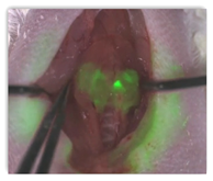
You can see in the video that the fluorescence was not contained only in the  cancer, but in adjacent areas as well. This is fascinating and explains how the pathology process works during surgery and she does a great job in describing how reports come back later, after surgery that find cancer tissue and then the treatment process continues from there with radiation.
Breast cancer and melanoma are discussed here as well and the surgeon looks for the first draining lymph node to determine where to cut. This process also provides what the doctor calls a “roadmap” for surgeons. The same smart technology can be used to find the cancer cells and tumor.
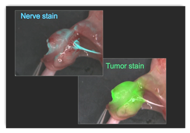
Swollen lymph nodes do not always mean cancer and this helps again direct the surgeon. She also mentioned nerve damage from surgery to include  prostate cancer surgery as that is a very small area for surgery. This is important as how many times do you read about “life” after prostate cancer surgery. They have found molecules they are now using to also find nerves.
When the two are put together the surgeon really appears to get even a much clearer road map and this does not say that pathology is gone by any means as that still remains part of the surgical procedures but doctors who have seen the process have commented that they feel this is positive for better potential surgical outcomes, after all this appears to be a great tool with identifying where the naked eye and other current technologies leave off. BD

